Regarding [#PetName], here is some information about Corneal Ulcer
Description:
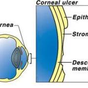
The cornea is the outermost transparent portion of the eye. It is made up of several (4) separate layers. The outermost layer of cells is called epithelium. The loss of these outermost cells results in an area of ulceration (corneal ulcer).
The cornea protects the eye from bacteria. Corneal ulcers left untreated can lead to blindness or loss of the eye.
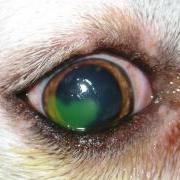
Causes:Corneal ulcers are usually caused by direct mechanical injury to the corneal surface. This can happen from sand, debris, plant material (grass or foxtail awns), thorns and scratches from other animals or objects.
Defects in the eyelids from eyelashes pointing inward against the cornea, or irritation from lack of tear production (dry eye) can cause ulcers.
Certain types of Herpes Viruses can cause corneal ulcers in affected cats.
Breeds with short snouts and bulging eyes (Boxers, Pugs, Pekinese etc.) are more prone to corneal injury. Boxers in particular are prone to a type of recurring ulcer known as indolent ulcer.
Indolent ulcers have a lip of damaged tissue around the periphery that slow or prevent the migration of healthy cells to repair the ulcer.
Symptoms:
Pets with corneal ulcers usually show the following symptoms:
Diagnosis:
Diagnosis is achieved by staining the eye with a mild dye called fluorescein. This will outline the size and perimeter of the ulcer and give an indication of how deep into the corneal layers it extends. Numbing the eye with a topical anesthetic may be necessary to encourage the pet to relax the eyelids for inspection of the cornea.
Treatment:
Healing occurs by the migration of new epithelial cells across the ulcerated area to replace the damaged and missing cells.
Naturally, eliminating the inciting cause is the first objective so that no further ulceration occurs. This most commonly involves removing the foreign debris from the eye and flushing with a sterile saline solution. Deep or indolent ulcers require debridement (surgical trimming) of damaged tissue or a procedure called a grid keratotomy to speed up the healing. A third eyelid flap surgery may be required to protect the eye during the healing process in the case of deep ulcers.
The second objective in treatment is the prevention of secondary infection. Oral antibiotics and antibiotic eye ointments accomplish this.
Pain, if present, can be managed with certain eye ointments (Atropine) and NSAID’s.
Warm compresses enhance blood supply and speed the rate of healing. Applying warm compresses (washcloth soaked in warm water) for 10 minutes twice daily will shorten healing time.
Superficial ulcers will heal in 5 to 7 days. Indolent and deep ulcers may take several weeks to heal.
During the healing process, you may notice blood vessels forming on the surface of the eye (neovascularization). This is initially good as the blood supply will bring nourishment and speed up the rate of repair. Once the ulcer is filled in, however, reducing the blood vessels is cosmetically important. Eye ointments with steroids are used to diminish the blood vessels. It is important not to use steroids in the eye until the ulcer is healed and no longer picking up dye when stained.
Other Information:
Close follow-up to monitor proper healing is especially important.

